텍스트를 입력해주세요
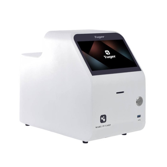
Tuger Fluorescence Cell Analyzer
Tuger is equipped with two fluorescent excitation wavelengths and two detection filters. It offers additionally a bright field view on all samples in the digital microscope optics. The analyzer allows for the routine analysis of cell density and viability, and transfection efficiency. The pre-configured apps guarantee an easy, anyhow safe execution of all tests.

Patented autoFocus automated cell analyzer
Tuger enables multi-channel and multi-field sampling. It provides automatic focusing of samples to ensure a clear image and accurate results. The use of light sources and filter wheel design greatly enhances system stability and operability.
Five fields of view per sample with high repeatability and fast image per processing
During the counting process, Tuger samples 5 fields of view per sample and simultaneously collects bright field and two fluorescence channel image information. The larger sample acquisition volume ensures counting accuracy. Tuger can complete fluorescence analysis for a single field of view within 4 seconds and provide fluorescence analysis results for 6 samples in just 2 minutes*.
*Results from a single fully automated test of six samples of suspension culture cells at a concentration of 1×10⁶cells/mL.


Multi-fluorescence synchronous detection
Tuger can provide up to 8 excitation lights and 6 filters, which allows for combinations of up to 15 fluorescence channels*.It fufills various cell application requirements. Set up your desired experimental protocols, then let Tuger take control of the experiment.
*Up to eight excitation light emitters are available(365nm、475nm、545nm、630nm)And six types of optical filters(445nm、530nm、553nm、605nm、635nm、695nm),Up to 15 combinations of fluorescence channels can be achieved.
FDA’s 21 CFR Part 11 complient operation
Tuger provides features such as multi-user levels and permissions management, encrypted data export, electronic signatures and records, audit trail, and more. These functionalities ensure accurate, reliable, and consistently stable results while also providing the capability to identify illegal or tampered records.

AO/PI Double fluorescent staining*

Cell transfection——GFP Fluorescence detection*

*The experimental images are from the Newton Optical Research Institute, and both liveness counting and cell transfection were performed using Hela cells.
| General | |
| Name | Tunin Cell Counter |
| Dimensions(W×D×H) | 245mm×139mm×205mm |
| Weight | 2.2kg(Excluding power adapter) |
| Screen | 7-inch multi touch color display screen |
| Camera | Industrial grade CMOS 6.3 million pixels (color high-definition) |
| Storage | Standard 128GB hard drive, other capacities are optional |
| Other Interfaces | 2 USB ports and 1 RJ45 network port |
| Applicable consumables | EasyFive six-digit counting slide |
| Technical parameters | |
| concentration range | 1×10⁴ - 3×10⁷ cells/mL |
| Diameter range | 5 - 200μm |
| Viability range | 0 - 100% |
| Sample volume | 10μL(GPC100,GR100)/20μL(GPC200,GR200) |
| Measurement time | <1s |
| Output format | jpg、PDF、csv |
| Features | 6-slot counting high-throughput, FDA, Lims, dilution calculator |
| Item number | |
| Tunin Cell Counter | E1TNX02 |
| Six cell counting slide (1 box, 50 pieces) | N3EF120(GPC200);N3EF110(GPC100) |
| Reusable counting slide (1 box, 1 piece) | N3EFR200(GR200);N3EFR100(GR100) |
| 0.2% Trypan Blue dye (5mL/bottle) | N3TP020 |